3D-Guided Surgery: Russian Surgeons Remove Jaw Tumor and “Print” a New Bone for a Patient
Oncologists in Chelyabinsk performed a procedure that combined virtual modeling, 3D printing, and highly precise surgical craftsmanship. A patient with a lower-jaw tumor received a chance to return to a full and active life.
Cancer is a frightening and life-altering diagnosis in its own right. The challenge becomes even greater when a tumor affects the face. In such cases, physicians face a dual objective: not only saving the patient's life but also preserving the ability to live, work, and interact socially without significant facial deformities.
In Chelyabinsk, a patient with cancer affecting the maxillofacial region required a complex surgical intervention. The tumor had invaded part of the lower jawbone, leaving a substantial defect already forming in the affected area. Simply covering the defect with a plate after tumor removal was not an option, because the patient would have lost the ability to breathe, eat, and speak normally. The surgical team therefore pursued another path: replacing the removed bone with a living tissue transplant. They achieved this through three-dimensional surgical planning.
Operation as a Construction Set
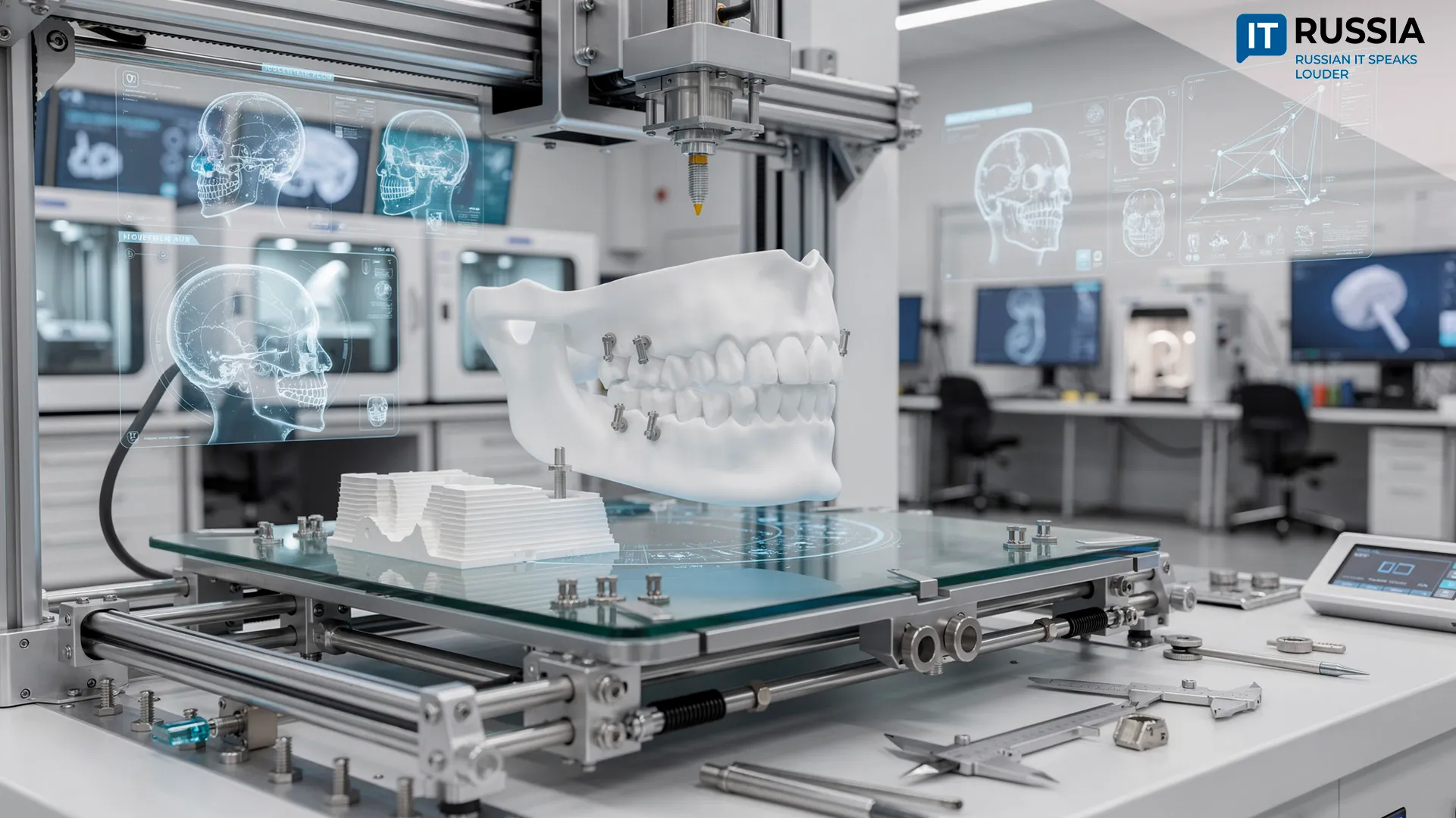
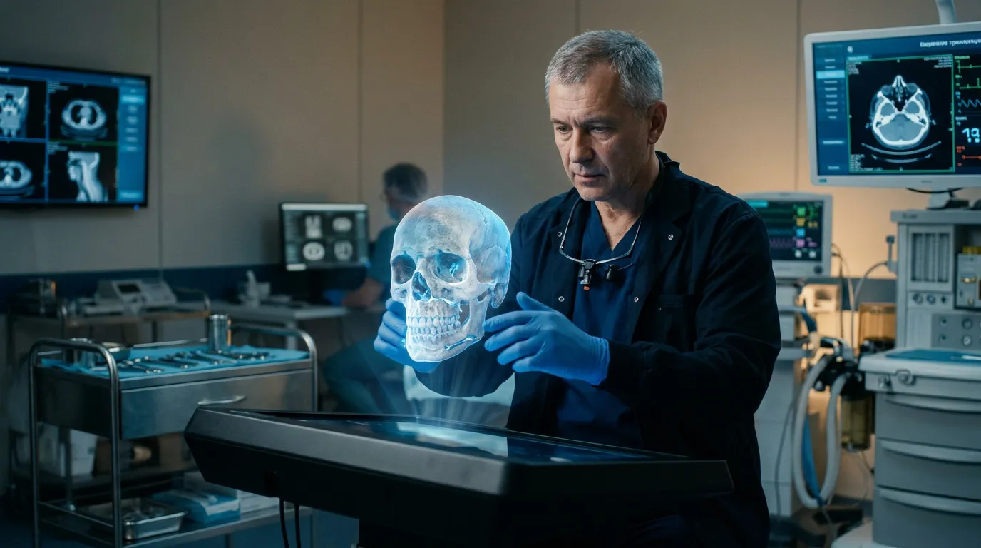
The process began with a CT scan. Using those images, specialists created an accurate 3D model of the patient's facial skeleton. Surgeons then rehearsed the procedure on the digital model in advance, determining exactly which section of the jaw needed to be removed, how a graft should be shaped from the patient's own fibula, and what type of titanium fixation plate would be required.
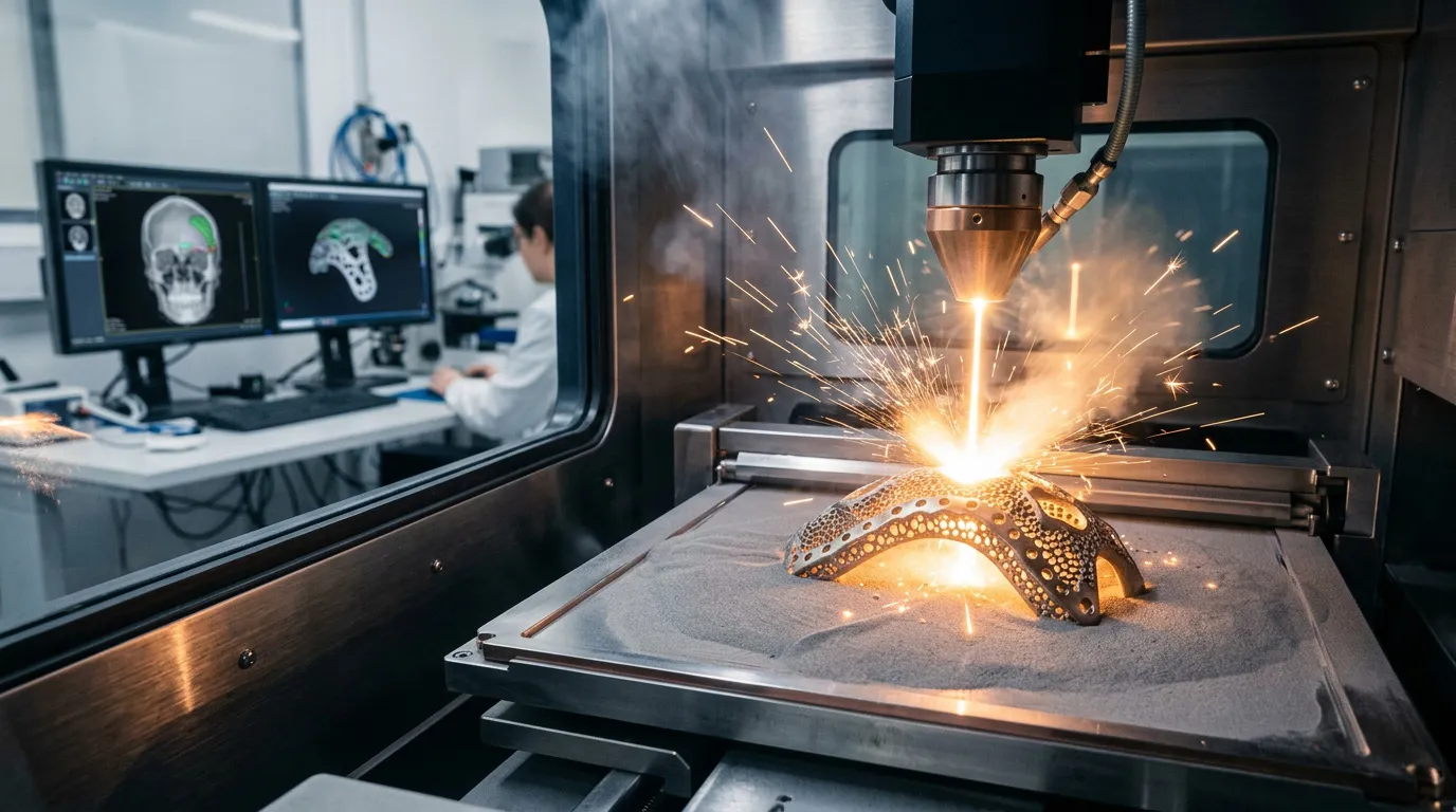
The most meticulous stage of the procedure involved work on the patient's fibula. Surgeons needed to remove a segment that would precisely match the shape and curvature of the missing section of the jaw. The cutting lines were calculated on the digital model with sub-millimeter precision. Based on those calculations, the team produced surgical guides and manufactured a patient-specific titanium plate using 3D-printing technology.
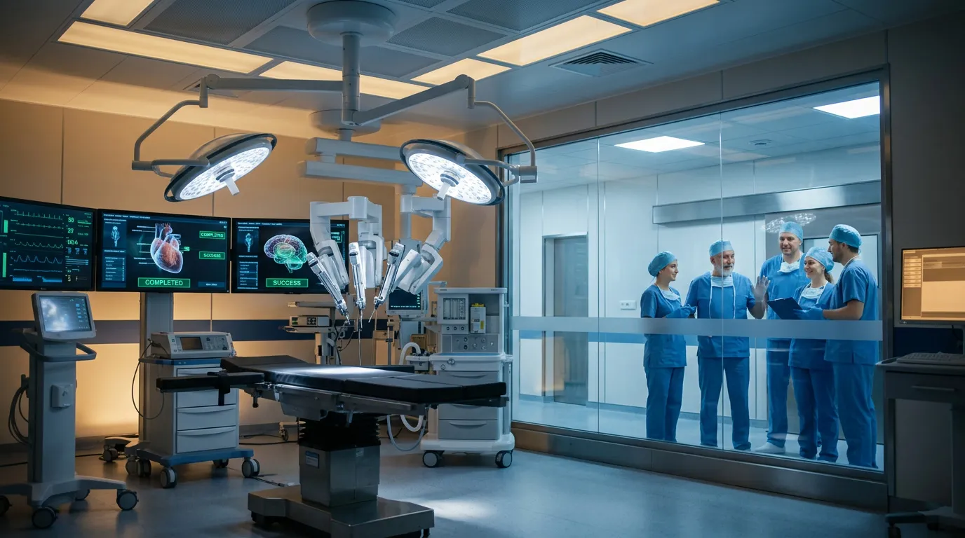
Seven Hours in the Operating Room
The highly complex operation lasted seven hours. Surgeons followed a carefully calculated plan: first removing the tumor, then harvesting a segment of lower-leg bone, shaping it into the missing portion of the jaw, and securing the reconstruction with the titanium plate. As a result, the components fit together without additional adjustments. That level of accuracy was made possible by the preoperative 3D model.
According to the surgical team, using the patient's own tissue for reconstruction reduces the risk of rejection, improves graft integration, and creates better conditions for full recovery. The patient has already been discharged without complications. He is able to walk independently and eat on his own, and he is expected to regain dental function in the future. Implant positions were incorporated into the reconstruction in advance. For an oncology patient, that represents a remarkable outcome.
A 3D Model for Physicians and Patients
For both physicians and patients, 3D modeling brings substantial advantages to surgery. Most importantly, it removes much of the uncertainty from the procedure. Surgeons know in advance what tissue must be removed, where replacement bone will come from, how it will be fixed in place, and what the final result is expected to look like.
The approach also reduces the risk of damaging nerves and blood vessels. Recovery times become shorter, patients return to normal life more quickly, and they avoid the psychological burden that can accompany visible facial defects. That benefit is no less important than the technical success of the operation itself.
What It Means for the Region and the Country
For the Chelyabinsk Region, the operation confirms that its regional oncology center can perform highly advanced reconstructive procedures without sending patients to Moscow or St. Petersburg. For Russia, it offers another notable example of personalized medicine expanding beyond federal centers and into regional healthcare systems. Just a few years ago, technologies of this kind were available at only a handful of major national institutions. Today they are being adopted in Chelyabinsk, and this is far from an isolated case.
The most significant domestic opportunity lies in expanding these practices across regional cancer centers, maxillofacial surgery departments, trauma units, and neurosurgical clinics. Three-dimensional modeling is especially valuable in cases where standard approaches are insufficient. These include head and neck cancers, jaw defects, skull injuries, complex joint-replacement procedures, and bone reconstruction following oncological disease.
As a result, Russia is advancing in parallel with the global shift toward personalized medicine.